Building Micro Synapses with Spinules
Your brain is made of billions of special cells called neurons that form over a trillion connections. These connections, called synapses, let you learn, create memories, and make you who you are.
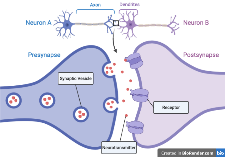
A synapse is where two neurons meet and transmit information. Neurotransmitters are released from the presynapse on Neuron A and bind to receptors on the postsynapse of Neuron B.
Neurons “talk” by making electrical pulses that travel along the neuron like a wire until they reach a synapse, where two neurons meet. When the electrical signal reaches the presynapse at the end of neuron A, small bubbles called synaptic vesicles dump chemicals called neurotransmitters into the space between the neurons. The neurotransmitters cross this gap and activate tiny switches called receptors on the dendrites of neuron B at the postsynapse. Dendrites look like tree branches, and dendritic spines, which hold the receptors, are the branches’ knobby leaves.

To see the neurons and spines, we make them glow with fluorescent dyes and proteins. This is a glowing neuron with many dendrites. The thorny looking “bumps” along the dendrites are the dendritic spines. Image by Christopher Pratt.
Dendritic spines are tiny; if you could mash them all together, 600 of them would still be smaller than a drop of water! But did you know that these dendritic spines can make even smaller synapses? A synapse within a synapse? I was part of a team that published some exciting new results that show some of these micro-synapses, called spinules (“tiny spines”), in action.
What the Heck are Spinules?
In the work led by Dr. Colleen Zaccard, we found that active dendritic spines can make tiny projections called spinules that reach out to neighboring neurons. Spinules have been observed before (Petralia et al., 2018; Spacek & Harris, 2004), but never in live, behaving cells! Spinules appear and disappear with most of them lasting less than 30 seconds, but some of them stick around for a while.

Spinules (arrows in A) are most abundant on mature mushroom spines (panel B), and 85% of observed mushroom spines had spinules (panel C). Adapted from Zaccard et al. (2020).
Dendritic spines come in a few shapes named after what they look like. As a synapse gets stronger, the spine becomes bigger and mushroom-shaped. Mushroom spines are the biggest, roundest, and most mature while filopodia, thin, and transitioning spines are less mature. Branched just means that there is more than one spine coming off the dendrite at the same place and is also typically still immature. As dendritic spines matured, they were more likely to have spinules, and 85% of the mature, mushroom dendritic spines had at least one spinule. If this is true for humans, then we all have these small synapses-on-synapses forming all the time!

Spinules (yellow) that persist a long time (more than 60 seconds) tend to be larger than those that don’t. On the bottom, you can see a long-lived spinule (blue arrow) compared to a short-lived spinule (red arrow) on a dendritic spine (red) over time. Adapted from Zaccard et al. (2020).
What are Spinules Doing?
This is the big question: We’re not quite sure. It’s difficult to do physiological studies on such small structures. We suspect that they are forming little synapses because they contain proteins that make postsynapses and they reach out and touch presynapses. These spinules also respond to neuronal activity, which means they could be a part of learning.

The green spots and arrows show postsynaptic density (PSD), an essential building block for synapses in the dendritic spines and spinules (red). 75% of long-lived spinules have this protein! Adapted from (Zaccard et al., 2020).
This just shows that as much as we think we know about how our bodies and biology work, there’s always more to be discovered! Classical neuroscience thinks of synapses as a one-to-one connection – one presynapse to one postsynapse, but this idea is evolving. Spinules might be a way that dendritic spines “explore” and build multiple connections, which could produce complex learning or associations. We also found that a gene involved in autism and schizophrenia controls spinules, so maybe these mysterious micro synapses do more than their size would suggest!

A spinule (red encircled with dashed white line) touching a distant presynapse (green). Adapted from (Zaccard et al., 2020).
The paper uncovered much more than what I’ve written about here. We used mouse cells and brains in our experiments, so the situation might be a little different in humans; still, it’s remarkable how similar brains are across the animal kingdom! In this study, I aided in experimental design (notably in labeling the presynapses in the last image in this post) and data analysis. For a limited time, you can find the original paper here , otherwise email me!
References
Arellano, J. I., Benavides-Piccione, R., DeFelipe, J., & Yuste, R. (2007). Ultrastructure of Dendritic Spines: Correlation Between Synaptic and Spine Morphologies. Frontiers in Neuroscience, 1(1), 131–143. https://doi.org/10.3389/neuro.01.1.1.010.2007
Measured average spine volume as 0.08 µm^3. A drop of water is roughly 50 µm^3.
Petralia, R. S., Wang, Y.-X., Mattson, M. P., & Yao, P. J. (2018). Invaginating Structures in Mammalian Synapses. Frontiers in Synaptic Neuroscience, 10. https://doi.org/10.3389/fnsyn.2018.00004
Spacek, J., & Harris, K. M. (2004). Trans-Endocytosis via Spinules in Adult Rat Hippocampus. Journal of Neuroscience, 24(17), 4233–4241. https://doi.org/10.1523/JNEUROSCI.0287-04.2004
Zaccard, C. R., Shapiro, L., Martin-de-Saavedra, M. D., Pratt, C., Myczek, K., Song, A., Forrest, M. P., & Penzes, P. (2020). Rapid 3D Enhanced Resolution Microscopy Reveals Diversity in Dendritic Spinule Dynamics, Regulation, and Function. Neuron, 107(20). https://doi.org/10.1016/j.neuron.2020.04.025